Institute for Advanced
Medical Education
Ultrasound-Focused CME for
Physicians and Sonographers

ACCME Accredited to Award
AMA PRA Category 1™ CME

ARDMS Sponsor
THE UNLIMITED CME PLAN
Unlimited 2024
-
75% less compared to competitors
-
Instant access to unlimited CME from our entire member's library of high quality ultrasound-focused CME. Fulfill all your CME requirements in one place, for one LOW price!
-
Unlimited CME for 2024
-
ACCME Accredited Courses
-
CME Credits Automatically Transfer
-
Membership Expires
12/31/2024
Lifetime CME
-
Save over $1200 on Unlimited CME for until the end of 2025
-
The best choice for CME focused on ultrasound for physicians!
-
Unlimited CME for life
-
Premium Courses
-
Exclusive Content
-
ACCME Accredited Courses
-
CME Credits Automatically Transfer to ACCME
Unlimited Plus
-
Save 40% today! The value of this product is $299.99
-
Instant Access Now for 2024 and All of 2025 to our Unlimited CME Plan. Get a head start on your CME today with the Unlimited Plus Plan!
-
Unlimited CME 2024-2025
-
ACCME Accredited Courses
-
CME Credits Automatically Transfer
-
Membership Expires
12/31/2025
All Memberships Include
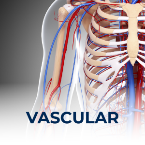
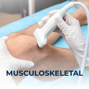
- Access to 150+ Ultrasound Focused Courses in 10 Specialties.
- Precision in hands-on technique showcased by experts
- Credits are automatically uploaded to your ARDMS bank and stored on your
- account to be accessed at any time. In the event of an audit, we've got your back.
Unlimited Access
Premier Content
Audit Protection
WHO ACCEPTS IAME CME


PROUD PARTNERS WITH


ARDMS CME AUDIT PROCESS
Every year the ARDMS conducts audits for the participants in the closing 3 year period. When conducting the audit the ARDMS must verify your courses werre completed by a reputible company and that the certificates are authentic. When the ARDMS see's your credits are from IAME, the audit process is complete because the IAME sends credits to the ARDMS electronically using propietary API technology that most competitors have not invested in. The Institute for Advanced Medical Education is your friend in the process working directly with the ARDMS
Course Library
Happy Customers

Lifetime
CME Membership
Continuous Access to Cutting-Edge Knowledge: At the Institute for Advanced Medical Education our Lifetime Membership grants you perpetual access to the latest in ultrasound education, keeping your skills sharp and current.

Premium Content
Our premium content goes beyond our traditional CME. Physicians, specialists, and advanced practice providers will find brand new content that is high-quality and high-yield. These training courses will cover topics in all specialties and all fields of medicine.
State-Required licensing courses will be available soon.


