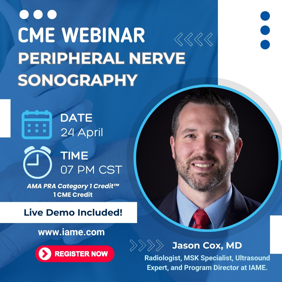
 |
|
INTRODUCTION |
Diagnostic
ultrasound is generally perceived by users and patients as a safe
technique with no adverse effects. Since ultrasound is so widely
used in pregnancy, it is essential for all practitioners to ensure
that its use remains safe. Ultrasound causes thermal and mechanical
effects in tissue which are increased as the output power is increased.
In
the last decade, there has been a general trend towards increased
output with the introduction of color flow imaging, more use of
pulsed ‘spectral Doppler’ and higher demands on B-mode imaging
1. In response to these increases, recommendations
for the safe use of ultrasound have been issued by several bodies.
In addition, recent regulations have changed the emphasis of responsibility
so that more onus is now placed on the operator to ensure that
ultrasound is used safely. This chapter summarizes the effects
and the standards issued and outlines recommendations for safe
use in obstetric practice.
|
EFFECTS |
Ultrasound
is a mechanical energy in which a pressure wave travels through
tissue. Reflection and scattering back to the transducer are used
to form the image. The physical effects of ultrasound are generally
categorized as:
(1)
Thermal effects – heating of tissue as ultrasound is absorbed by
tissue. Heat is also produced at the transducer surface;
(2)
Cavitation – the formation of gas bubbles at high negative pressure;
(3)
Other mechanical effects – radiation forces leading to streaming
in fluids and stress at tissue interfaces.
The
implications of these effects have been determined by in vitro,
animal and human epidemiological studies and are briefly summarized
below. |
Thermal
effects |
As
the ultrasound waves are absorbed, their energy is converted into
heat. The level of conversion is highest in tissue with a high absorption
coefficient, particularly in bone, and is low where there is little
absorption (e.g. amniotic fluid). The temperature rise is also dependent
on the thermal characteristics of the tissue (conduction of heat
and perfusion), the ultrasound intensity and the length of time
for which the tissue volume is scanned. The intensity is, in turn,
dependent on the power output and the position of the tissue in
the beam profile. The intensity at a particular point is altered
by many of the operator controls, for example power output, mode
(B-mode, color flow, spectral Doppler), scan depth, focus, zoom
and area of color flow imaging. With so many variables, it has proved
difficult to model temperature rises in tissue. In
vitro studies have been used with a ‘worst case’ model of tissue
to predict temperature rises o, for instance in the formation
of thermal indices (see below). The transducer face itself
can become heated during an examination. Heat is localized to the
tissue in contact with the transducer. |
Cavitation |
Cavitation
is the formation of transient or stable bubbles, described as inertial
or non-inertial cavitation. Inertial cavitation has the most potential
to damage tissue and occurs when a gas-filled cavity grows, during
pressure rarefaction of the ultrasound pulse, and contracts, during
the compression phase. Collapse of the bubble can generate local
high temperatures and pressures.
It
has been hypothesized that ultrasonically induced cavita tion is
the cause of hemorrhage in the lungs and intestines in animal studies
2–6 . In these studies, effects have been seen at tissue
interfaces with gas. The absence of gas in fetuses means that the
threshold for cavitation is high and does not occur at current levels
of diagnostic ultrasound. The introduction of contrast agents leads
to the formation of microbubbles that potentially provide gas nuclei
for cavitation. The use of contrast agents lowers the threshold
at which cavitation occurs, but this is not current practice in
obstetrics. |
|
Other
mechanical effects |
The
passage of ultrasound through tissue causes a low-level radiation
force on the tissue. This force produces a pressure in the direction
of the beam and away from the transducer and should not be confused
with the oscillatory pressure of the ultrasound itself. The pressure
that results and the pressure gradient across the beam are very
low, even for intensities at the higher end of the diagnostic range
7. The effect of the force is manifest in volumes of
fluid where streaming can occur with motion within the fluid.
The fluid velocities which result are low and are unlikely to cause
damage. |
|
Effects
on fetuses |
Effects
are divided into mechanical and thermal. For mechanical effects,
there is no evi-dence that cavitation occurs in fetal scanning.
In a study of low-amplitude lithotripsy pulses in mouse fetuses,
there has been concern that hemorrhage may be the result of tissue
movement caused by radiation forces 8 . There is no evidence
that this occurs in vivo in fetal scanning. The primary concern
in fetal imaging is temperature rise. It is known that hyperthermia
is teratogenic. The efforts of investigators have concentrated on
defining the temperature increases and exposure times which may
give rise to biological effects and on determining the ultrasound
levels which might, in turn, lead to those temperature rises. With
this information, criteria have been identified for the safe use
of diagnostic ultrasound.
Temperature
rises of 2.5°C have been demonstrated in excised unperfused guinea
pig brain tissue after 2 minutes’ exposure to ultrasound at the
high end of pulsed wave Doppler ultrasound intensity levels 9
. At the bone surface, temperature increases of up to 5°C were found.
In a study on sheep using different intensity criteria 10
, the temperature rise in utero was found to be 40% lower
than that in the equivalent non-perfused test. While the observed
temperature increases occurred in high-intensity modes (typical
of pulsed wave Doppler used at maximum power), these levels of intensity
are achievable with some current scanner/transducer combinations.
The
issue of sensitivity of fetal tissue to temperature rise is complex
and is not completely understood. Acute and chronic temperature
rises have been investigated in animals, but study designs and results
are varied. Work carried out in this field is summarized elsewhere
11 .
The
uncertainty over chronic changes is reflected in the WFUMB guidelines
12 . These state that ultrasound that produces temperature
rises of less than 1.5°C may be used without reservation. They also
state that ultrasound exposure causing temperature rises of greater
than 4°C for over 5 min should be considered potentially hazardous.
This leaves a wide range of temperature increases which are within
the capability of diagnostic ultrasound equipment to produce and
for which no time limits are recommended. |
|
Epidemiology |
Several
studies have examined the development of fetuses receiving different
levels of ultrasound investigation. In trials comparing ultrasound
screened and non-screened groups, there has generally been no difference
in birth weights between groups. There have been no unequivocal
data to suggest that there is impaired development of hearing, vision,
behavior or neurological function due to ultrasound screening. In
a large, randomized trial of over 3200 pregnant women in which half
were offered routine ultrasonography at 19 and 32 weeks, there was
no evidence of impaired growth or neurological development up to
follow-up at 8–9 years. There was a possible association of left-handedness
amongst boys undergoing ultrasonography 13
. Scanning of this group was performed with B-mode only. There have
been concerns that epidemiological studies to date do not reflect
the higher output capabilities of modern scanners. |
|
OUTPUT
REGULATIONS, STANDARDS AND GUIDELINES
– WHO DOES WHAT? |
Regulations
governing the output of diagnostic ultrasound have been largely
set by the USA’s Food and Drug Administration (FDA), although the
International Electrotechnical Commission (IEC) is currently in
the process of setting internationally agreed standards.
The
relevant national societies for ultrasound users (e.g. American
Institutue of Ultrasound in Medicine (AIUM), British Medical Ultrasound
Society (BMUS)) usually have safety committees who offer advice
on the safe use of ultrasound. In 1992, the AIUM, in conjunction
with the National Electrical Manufacturers Association (NEMA) developed
the Output Display Standard (ODS), including the thermal index and
mechanical index which have been incorporated in the FDA’s new regulations
14,15.
Within
Europe, the Federation of Societies of Ultrasound in Medicine and
Biology (EFSUMB) also addresses safety and has produced safety guidelines
(through the European Committee for Ultrasound Radiation Safety).
The World Federation (WFUMB) held safety symposia in 1991 (on thermal
issues) and 1996 (thermal and non-thermal issues), at which recommendations
were proffered. Following review, these were published in 1992 and
1998 as guidelines. |
|
Past
regulations |
The
initial FDA regulations on ultrasound output were produced in 1976.
These imposed application-specific limits, based on existing output
levels which had demonstrated no adverse effects. Limits were divided
into:
(1)
Ophthalmic applications;
(2)
Fetal and other (including abdominal, pediatric, small parts);
(3)
Cardiac;
(4)
Peripheral vessels.
For
spatial peak time-averaged intensity (I-SPTA) (the measure most
associated with temperature rise), the maximum levels were:
Ophthalmic |
17
mW/cm 2 |
Fetal
and other |
94
mW/cm 2 |
Cardiac |
430
mW/cm 2 |
Peripheral
vessel |
720
mW/cm 2 |
Scanners
typically had a key/button which limited output for obstetric applications.
Although power and intensity limits could be exceeded in some scanners,
especially when using pulsed wave Doppler or color Doppler, this
required a deliberate effort on the behalf of the users. |
|
Current
regulations |
In
revising its regulations in 1993, the FDA 15
altered its approach to ultrasound safety. The new regulations combine
an overall limit of I-SPTA of 720 mW/cm 2 for all equipment with
a system of output displays to allow users to employ effective and
judicious levels of ultrasound appropriate to the examination undertaken.
The new regulations allow an eight-fold increase in ultrasound intensity
to be used in fetal examinations. They place considerably more responsibility
on the user to understand the output measurements and to use them
in their scanning. The output display is based on two indices, the
mechanical index (MI) and the thermal index (TI). |
|
Mechanical
index |
The
mechanical index is an estimate of the maximum amplitude of the
pressure pulse in tissue. It gives an indication as to the relative
risk of mechanical effects (streaming and cavitation). The FDA regulations
allow a mechanical index of up to 1.9 to be used for all applications
except ophthalmic (maximum 0.23). |
|
Thermal
index |
The
thermal index is the ratio of the power used to that required to
cause a maximum temperature increase of 1°C.
A thermal index of 1 indicates a power causing a temperature increase
of 1°C. A thermal index of 2 would be twice that power but would
not necessarily indicate a peak temperature rise of 2°C. Because
temperature rise is dependent on tissue type and is particularly
dependent on the presence of bone, the thermal index is subdivided
into three indices:
(1)
TIS: thermal index for soft tissue;
(2)
TIB: thermal index with bone at/near the focus;
(3)
TIC: thermal index with bone at the surface (e.g. cranial examination).
For
fetal scanning, the highest temperature increase would be expected
to occur at bone and TIB would give the ‘worst case’ conditions.
The mechanical index and thermal index must be displayed if the
ultrasound system is capable of exceeding an index of 1. The displayed
indices are based on the manufacturer’s experimental and modelled
data. These measurements are not infallible; an independent study
has demonstrated significant discrepancies over declared I-SPTA
output of up to 400% 16. |
|
Future
IEC standards |
An IEC standard (Draft IEC 61681) is being drawn up to establish
a safety classification for ultrasound equipment based on its ability
to produce cavitation or a temperature rise. The standard proposes
two classifications of equipment: class A, which has a lower output
and for which no output display is required, and class B which has
a higher output and for which an output display is required. The
draft is currently undergoing review. |
|
Guidelines |
Ultrasound
organizations have produced statements on the safe use of ultrasound.
These are not regulatory statements but are intended to educate
and advise. WFUMB guidelines have been issued in two special issues
of Ultrasound
in Medicine and Biology 12-17 . Statements and recommendations
are given on B-mode scanning, Doppler imaging, transducer heating,
thermal effects (see page 33). The AIUM have produced statements
on the safety of ultrasound. They are available from the AIUM office
and can be obtained from the AIUMwebsite – http://www.aium.org/stmts.htm.
The European Committee for Ultrasound Radiation Safety has published
statements 18,19 on the use of pulsed Doppler measurement in fetuses,
stating that its use in routine examinations during the period of
organogenesis is considered inadvisable at present. |
|
A
PRACTICAL APPROACH TO SAFE FETAL SCANNING |
No injurious effects
have been identified from ultrasound scanning of the fetus. However,
changes in power output, increased use of Doppler ultrasound and
a change in regulations governing outputs means that every measure
should be taken by users to maintain safe practices. |
Scanning
practice |
-
The ALARA ("As Low As
Reasonably Achievable")
principle should be maintained. Power outputs used should be
adequate to conduct the examination. If in doubt, use a low
power and increase it as necessary. Application keys for obstetrics
should bring in each mode at its lowest output so that the operator
is required to increase power if the examination demands it.
-
B-mode generally has the lowest power output and intensity.
M-mode, color flow and spectral Doppler have higher outputs
which can cause more heating at the site of examination. The
examination should begin with B-mode and use color and spectral
Doppler only when necessary.
-
The
intensity (and temperature rise) is highly dependent on scanner
settings.
For example, the intensity changes in response to changes in:
(a) Power Output,
(b)
Depth of examination,
(c)
Mode used (color flow, spectral Doppler),
(d)
Transmitted frequency used,
(e)
Color pulse repetition frequency (scale),
(f)
Region of color flow interest,
(g)
Focus.
- If
the display for the scanner/transducer combination shows thermal
and mechanical indices, the indices should be readily visible.
Of the thermal indices, TIB is most relevant to heating in the
second and third trimesters. The operator should be aware of changes
to the indices in response to changes in control settings.
- Special
care should be taken in febrile patients, since ultrasound heating
will cause additional heating to the fetus.
- The
WFUMB recommends that ultrasound causing a temperature rise of
no more than 1.5°C may be used without reservation on thermal
grounds.
- Thermal
indices exceeding 1.5 should not be used routinely and, if required
for specific diagnostic information, should be used for the minimum
time necessary. The influence of higher intensity levels can be
moderated by moving the transducer so that specific areas of tissue
are not subjected to long periods of higher intensity investigation.
- Do
not scan for longer than is necessary to obtain the diagnostic
information.
|
|
SELECTED
WFUMB STATEMENTS ON THE SAFETY OF DIAGNOSTIC ULTRASOUND |
|
B-mode
imaging (issued 1992) |
Known
diagnostic ultrasound equipment as used today for simple B-mode
imag- ing operates at acoustic outputs that are not capable of producing
harmful temperature rises. Its use in medicine is therefore not
contraindicated on thermal grounds. This includes endoscopic, transvaginal
and transcutaneous applications. |
|
Doppler
(1992) |
It
has been demonstrated in experiments with unperfused tissue that
some Doppler diagnostic equipment has the potential to produce biologically
significant temperature rises, specifically at bone/soft tissue
interfaces. The effects of elevated temperatures may be minimized
by keeping the time during which the beam passes through any one
point in tissue as short as possible. Where output power can be
controlled, the lowest available power level consistent with obtaining
the desired diagnostic information should be used.
Although
the data on humans are sparse, it is clear from animal studies that
exposures resulting in temperatures less than 38.5°C can be used
without reservation on thermal grounds. This includes obstetric
applications. |
|
Transducer
heating (1992) |
A substantial
source of heating may be the transducer itself. Tissue heating from
this source is localized to the volume in contact with the transducer. |
|
Recommendations
on thermal effects (1997) |
A
diagnostic exposure that produces a maximum temperature rise of
no more than 1.5°C above normal physiological levels (37°C) may
be used without reservation on thermal grounds.
A
diagnostic exposure that elevates embryonic and fetal in situ
temperature to 4°C (4°C above normal temperature) for 5 min
or more should be considered potentially hazardous. |
|
REFERENCES |
1.
Henderson J, Willson K, Jago JR, et
al. A survey of the acoustic outputs of diagnostic ultrasound equipment in current
clinical use in the Northern Region. Ultrasound Med Biol
1995;21:699–705.
2. Baggs R, Penney DP, Cox C, Child SZ. Thresholds for ultrasonically induced
lung hemorrhage in neonatal swine. Ultrasound Med Biol
1996;22:119–28.
3. Dalecki D, Child SZ, Raeman CH, Cox C, Carstensen EL. Ultrasonically-induced
lung haemorrhage in young swine. Ultrasound Med Biol
1997;23:777–81.
4. Frizzell LA, Chen E, Chong L. Effects of pulsed ultrasound on the mouse neonate:
hind limb paralysis and lung haemorrhage. Ultrasound Med Biol
1994;20:53–63.
5. Holland CK, Zheng X, Apfel RE, Alderman JL, Fernandez L, Taylor KJW. Direct
evidence of cavitation in vivo from diagnostic ultrasound. Ultrasound Med Biol
1996;22:917–25.
6. Zacchary JG, O’Brien WD. Lung lesions induced by continuous and pulsed wave
(diagnostic) ultrasound in mice, rabbits and pigs. Vet Pathol 1995;32:43–54.
7. Duck FA. Acoustic streaming and radiation pressure in diagnostic applications:
what are the implications? In Barnett SB, Kossoff G, eds. Safety of Diagnostic Ultrasound. Carnforth, UK: Parthenon
Publishing, 1998:87–98.
8. Dalecki D, Child SZ, Raeman CH, Penney DP, Mayer R, Cox C, Carstensen EL.
Thresholds for fetal haemorrhages produced by a piezoelectric lithotripter.
Ultrasound Med Biol 1997;23:287–97.
9. Bosward KL, Barnett SB, Wood AKW, Edwards MJ, Kossoff G. Heating of the guinea
pig fetal brain during exposure to pulsed ultrasound. Ultrasound Med Biol 1993;19:415–24.
10. Duggan PM, Liggins GC, Barnett SB. Ultrasonic heating of the brain of the
fetal sheep in utero. Ultrasound Med Biol 1995;21:553–60.
11. Tarantal AF. Effects of ultrasound exposure on fetal development in animal
models. In Barnett SB, Kossoff G, eds. Safety of Diagnostic
Ultrasound. Carnforth, UK: Parthenon Publishing,
1998:39–51.
12.
Barnett SB, ed. Conclusions and recommendations on thermal and non-thermal mechanisms
for biological effects of ultrasound. In WFUMBsymposium on
Safety of Ultrasound in Medicine. Ultrasound Med Biol, 1998;24, special issue 13. Salveson
K, Vatten L, Eik-Nes S, Hugdahl K, Bakketeig L. Routine ultrasonography
in utero and subsequent handedness
and neurological development. Br Med J 1993;307:159–64.
14. AIUM / NEMA. Standard for Real-Time Display
of Thermal and Mechanical Acoustic Output Indices on Diagnostic
Ultrasound Equipment. Rockville: American Institute of Ultrasound in Medicine,
1992 15. FDA. Revised 510(k) Diagnostic Ultrasound
Guidance for 1993. Rockville, MD: Food and Drug Administration,
Center for Devices and Radiological Health, 1993.
16. Jago JR, Henderson J, Whittingham TA, Willson K. How reliable are manufacturer’s
reported acoustic output data? Ultrasound Med Biol
1995;12:135–6.
17. Barnett SB, Kossoff G. eds. Issues and recommendations regarding thermal
mechanisms for biological effects of ultrasound. In WFUMB Symposium on Safety and Standardization in Medical Ultrasound. Ultrasound Med Biol
1992;18, special issue.
18. European Federation of Societies for Ultrasound in Medicine and Biology.
Guidelines for the safe use of Doppler ultrasound
for clinical applications. Report from the European Committee
for Ultrasound Radiation Safety. Eur J Ultrasound
1995;2:167–8.
19. European Federation of Societies for Ultrasound in Medicine and Biology.
Clinical safety statement for diagnostic ultrasound. Report from the European Committee for Ultrasound Radiation
Safety. Eur J Ultrasound 1996;3:283. |
|